Introduction. Suture granuloma is uncommon complication of surgical procedures developing secondary to the use of non-absorbable sutures [1]. It is a foreign body type of granulomatous reaction that includes multinucleated giant cell formation. The development of suture granuloma is a chronic process [2].The overall incidence of granuloma is 1 in 1000-18000 procedures [3]. The incidence of granuloma reaction due to iatrogenic factors increases because of widespread use of novel surgical materials [3]. In this study, a case with silk suture granuloma that developed after high orchiectomy was reported.
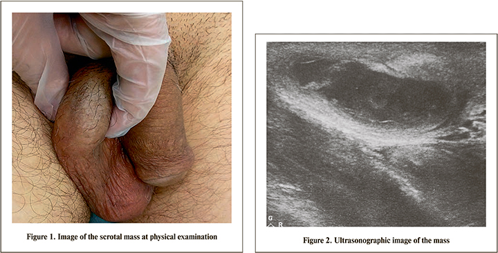
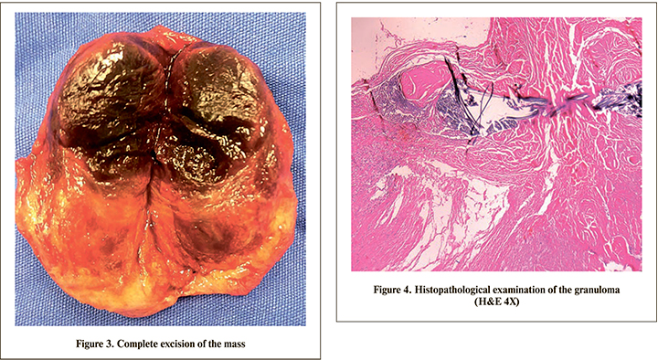
Case. A 59 year old man presented to clinic of urology, with one month history of painless scrotal swelling. In phsyical examination, the right testis was firm without enlarged lympadenopathy. Serum tumor markers were with normal range. The patient was treated with high orchiectomy. The histopathological diagnosis was granulomatous inflammation of the epididymes. After 3 weeks from the operation the patient applied to our clinic with 3*2 cm sized palpable, firm painless nodular mass in the right scrotum (Picture 1). Ultrasonography showed the palpable mass in the scrotum with a suggestion of granuloma (Picture 2). The patient was operated and the mass was excisionally removed (Picture 3) The histopathological examination reported the diagnosis of silk granuloma (Picture 4). The patient has no complaint during the three months follow up period.
Discussion. Suture granuloma is uncommon postoperative complication of the surgery devoloping from the nonabsorbable suture material [1]. It has two step process including the initial reaction of tissue inflicted the passage of the needle and specific inflammatory reaction of the suture material. Histologically, suture granuloma is a benign lesion that is characterized by a histiocytic reaction with foreign body giant cells around the sutures [2]. Developing suture granuloma may occur anywhere in the body and the risk of granuloma formation is much more in the use of silk and polyfilament materials than the absorbable sutures [1]. Silk sutures are preferred because of low cost, easy sterilization and stronger wound closure. Clinical presentation of the patients are; erythema, swelling, pain and solid mass [2]. The patient was presented with painless and progresively growing solid mass after the surgery in the present study. The time duration of the granuloma formation takes aproximetely from few months to several years [1]]. Seçil et al. [1], reported a case of suture granuloma in inguinal area 8 months after orchiectomy. In another study, the authors reported granuloma formation after 15 years from the surgery [4]. The occurence of granuloma is important for the patients who was diagnosed of any cancer. The lesions were confused with cancer recurrence and the patients were treated with unnecessary surgeries [5]. There are some diagnostic imaging modalities for suture granulomas. Ultrasonography is the most helpful imaging for the diagnosis of suture granuloma [1]. In general, hypoechoic lesion with/without posterior acustic shadowing and without doppler flow signal is usually seen in sonographic examination [2]. In superficial lesions; suture can be detected with high resolution devices [1]. Suture granulomas mimick intrabdominal tumors on computed tomography(CT) and PET/CT imagings. Suture granuloma appears hyperintense against a hypointense signal on magnetic resonance imaging technique [3]. In the present study, ultrasonography showed the superfical lesion that was suggested granuloma formation. The diagnosis of suture granuloma is surgical excision and histopathological examination [1] . Surgery is used to achieve both the diagnosis and eradication of the inflammation. Fine needle aspiration biopsy can be alternative for these patients [4].
Conclusion. The suture granulomas are very rare lesions in urology practice. The clinicans should be kept in mind that granuloma formation can be seen after surgery. The diagnosis needs histological examination with total excision or biopsy.